
Estimated reading time: 5 minutes
Magnetic resonance imaging (MRI) is a type of diagnostic test that produces cross-section three-dimensional imagery of the human anatomy. MRIs enables specialists to see through layers of organs through magnetism. It is a safer alternative to computed tomography (CT) scans and X-rays that use harmful radiation. Doctors and scientists employ the use of MRIs to observe the internal structure of the body to detect the following:
- Brain and spine abnormalities
- Specific heart issues
- Tumors and cysts anywhere in the body
- Anomalies in the abdominal organs
- Injuries and diseases
- Detecting causes of pelvic pain
- Evaluation of infertility
- Detecting issues at the joints and bones
The Ezra MRI scanner allows the early detection of serious diseases such as cancer, dementia, stroke, multiple sclerosis (MS), and more. These scanners can detect cancer in up to 14 organs in a span of an hour. Early detection of such anomalies raises the survival rate of patients through comprehensive reports. Definitely, annual MRI testing can help save people’s lives.
How Does MRI Work?
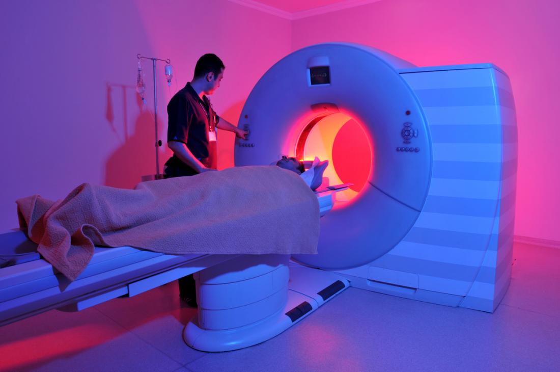
A patient is placed onto the patient table, which then automatically moves into a horizontal tube. The tube scanner has giant magnets that produce a significant magnetic field that aligns the hydrogen atoms in your body.
The protons within the hydrogen atoms are exposed to radio waves as well and disrupt them. The protons then give off a subtle signal that the sensor of the MRI scanner detects. It finally enables the linked computer to generate high-detailed black and white images.
MRI scanners can produce these images in slices. The technologist can manipulate the viewing at the bottom, front, and sides (axial, coronal, and sagittal). It further helps doctors to make a closer examination of the specific area in the body.
In some cases, agents such as gadolinium dye are used to further enhanced the images. The magnetic substance is injected into the bloodstream, absorbed by certain tissues, and stands out in the 3D image. Gadolinium thus enhances the images of veins and arteries as the scanner cannot detect moving fluid such as blood.
Specialists familiar with the body’s anatomical structure can then decipher its changes or anomalies through the imagery.
What the MRI Displays
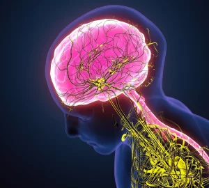
Magnetic resonance imaging can feature high-definition images of soft tissues such as the brain. Spinal fluid, blood, and bone marrow can vary from white to black. The presence of fat and water also affect the shades of white and black. Meanwhile, air and solid mass like bone appear black as they don’t give off signals. How these colors are distributed among the imagery is what the technologists try to decipher to determine the tissues’ health.
A spinal MRI can reveal much of the condition of the spine. If you experience difficulty breathing or coughing after suffering a spinal injury, it is considered an emergency case that you should not ignore, and you will almost certainly undergo an MRI to diagnose the cause. An MRI can detect pinched nerves, herniated discs, a fractured vertebra, a tumor, or compression.
An MRI of the neck and head can reveal a traumatic head injury, infection, headache causes, developmental issues, dementia, and more.
Scanning the arteries and veins can detect blockages, aneurysms, malformations, and carotid artery disease.
Preparation, During, and After an MRI Testing
The MRI scanner can easily detect underlying conditions when patients can do proper preparation. They will be asked to change into a gown.
There must not be any metallic objects in or on the body of the patient, so you should remove jewelry and accessories so that it won’t affect the scanner. It also means that those with irremovable metal objects such as old models of pacemakers cannot get an MRI.
If you have fillings or braces, they can distort the images of the MRI scanner. Your doctor and technologist will discuss this with you before the testing.
If you’re pregnant, you may also need to inform your doctor first. Doctors don’t recommend pregnant women at their first trimester to undergo MRI, but it is safe during the second and third trimesters.
Some patients may not be comfortable in enclosed spaces like the MRI scanner. In this case, it’s important to speak up and let the technologist know. You must be calm inside the scanner so you can go through the length of the process smoothly. You must relax and breathe in normally. The technologist will talk to you and make sure that you are ready.
Sometimes, specialists will inject an intravenous contrast liquid to make a specific tissue stand out on the computer screen. The patient must stay still so that the scanner can create clear images. You may receive instructions from your technologist, such as holding your breath, if necessary during the process.
If you suffered from a spinal injury, an MRI diagnostic would help determine the right treatment. The spine can have many issues, and treatment will depend on what the scanner presents. It will help your doctor decide what type of medication you need and if you need surgery. There are times that the spine may need braces to prevent damaging nearby soft tissues.
You may occasionally hear constant clicking noises related to the testing. Depending on the test’s complexity, it may last from 30–90 minutes.
After the scanning, the technologist will need to examine the images. Depending on the first images came out, you may need to undergo another MRI scanning. Once the scanning is complete, you will be sent home, and the technologist shall prepare a report for your doctor to discuss with you.
In Conclusion
MRI testing is a safer alternative to CT scans and X-rays because, unlike these other methods, it doesn’t use radiation. It instead uses magnetism to produce images of your internal organs. Your doctor will suggest this testing if necessary. Often those who have a brain, spinal, and arterial concerns are suggested to undergo the testing. Liquid agents may also be injected into your body to make specific tissues appear clearer. While the technology is complicated and useful, the patient must follow instructions so that the technologist can run a smooth diagnostic. This diagnostic tool is an absolute lifesaver for detecting diseases before it’s too late.

